Cardiac tamponade is caused by the accumulation of fluid in the pericardial space. If this occurs quickly, pressure rises in this space because the pericardium cannot stretch. If the pressure is higher than in the various chambers of your heart, they get compressed and some of the blood returning from your body gets backed up. This results in acute failure of the heart’s ability to pump blood back into your circulation.
Cardiac tamponade is a medical emergency.
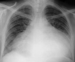
This Chest X Ray show a massive bottle shaped cardiac silhouet, caused by a large fluid collection around the heart.
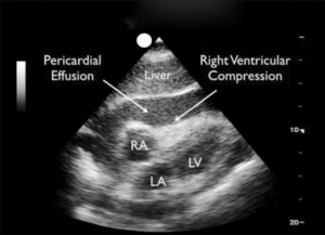
Cardiac ECHO shows a distended right atrium (RA), while both right (RV) and left (LV) ventricles (arrows) are compressed by the high pressures in the pericardial space, thus preventing blood from entering
Causes
Cardiac tamponade may occur as a complication of many different causes:
- Infection – Viral (HIV), bacterial (tuberculosis), fungal
- After various cardiac procedures such PCI and Heart Surgery
- Trauma to the chest
- Myocardial infarction
- Connective tissue diseases (Systemic lupus erythematosus, rheumatoid arthritis)
- Radiation therapy to the chest
- Renal failure
- Anticoagulation treatment
- Aortic Dissection
Symptoms
- Low blood pressure
- Rapid heart beats (Tachycardia)
- Distended neck veins, worse with inspiration (Kussmaul sign)
- Diminished heart sounds (Beck’s Triad: a combination of distended neck veins, hypotension, and diminished heart sounds)
- Cold, clammy extremities
- Anxiety, restlessness
Diagnosis
- Blood tests
- EKG, Chest X-ray
- Cardiac ECHO
Treatment
- Oxygen
- IV fluids
- Medications to support the blood pressure
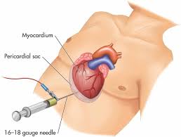
- Pericardiocenthesis
- Surgical exploration
- Correction of the underlying cause (if possible)


Comments 1
Pingback: Pericarditis and pericardial effusion - Cardiac Health